A virtual three-dimensional trips inside someone else’s head is now possible after scientists used new techniques to produce the most accurate images of the human brain ever revealed.
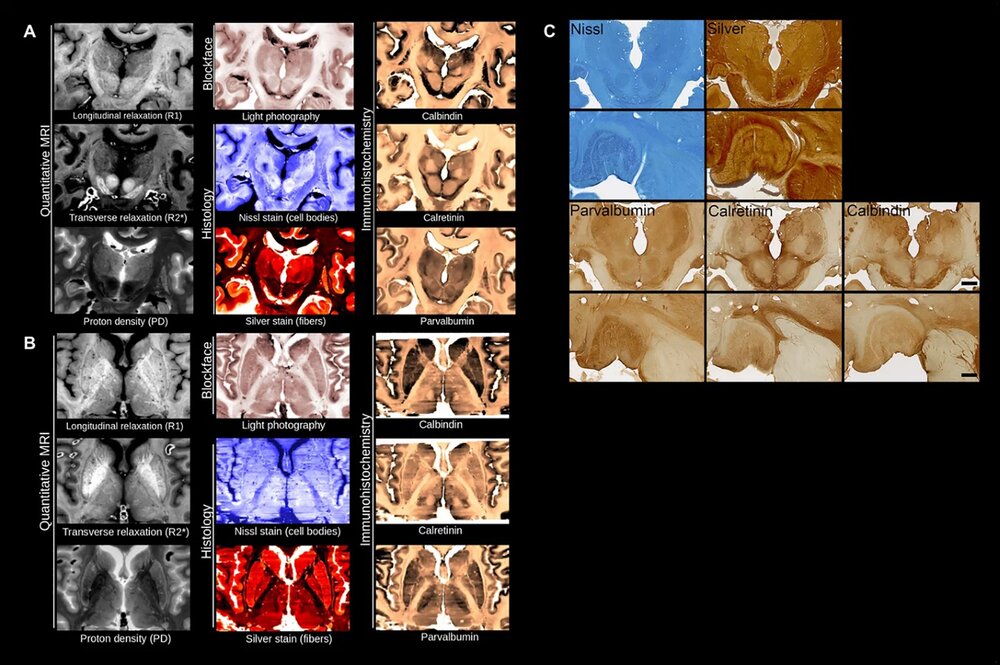
By combining non-invasive magnetic resonance imaging (MRI) and microscopes, a team of neuroantomists from the University of Amsterdam and the Max Planck Institute produced 3D images of two complete brains with an unprecedented level of detail.
Team member Anneke Alkemade, who is co-author of a study on the technique published in the journal Science Advances, said: “We are excited about all the possibilities this can open up for the field. Instructors can use the datasets for neuroanatomy training or virtual dissection, for example.”
She added, “And the ability to compare MRI results to individual proteins will give researchers more insight into poorly understood MRI observations and provide more anatomical details about small brain structures.
Co-author Nikolaus Weiskopf said: “It allows us to better link the MRI images to the underlying biological microstructures,”

He also added that the technique may detect dangerous medical conditions, such as Parkinson’s disease, much earlier than currently possible.
Co-author author Evgeniya Kirilina, said, “It’s a unique resource for studying very small but very important brain nuclei that supply the brain with the neurotransmitter dopamine.”
Dopamine is an organic molecule that plays several important roles in cells, including in the brain. In the brain, dopamine is a neurotransmitter—a chemical released by nerve cells to send signals to other nerve cells.
In the brain, there are several dopamine pathways, of which one has an important role in reward-motivated behaviour.
The level of dopamine in the brain increases in an anticipation of rewards, while many addictive drugs increase dopamine release or block its reuptake into neurons.
In addition, there are nervous system diseases associated with dysfunctions of dopamine, including Parkinson’s disease, attention deficit disorder (ADHD), restless legs syndrome, and schizophrenia.
In the new study, the researchers used an MRI much more powerful than the hospital version, relying on software they developed to distinguish between living and preserved human tissue.

Sectioning brain tissues into thin slices, the team photographed each section individually and digitally corrected them later.
Each section was placed on special glass slides and processed on custom-built laboratory equipment.
After the sections were digitized, the co-authors created new algorithms to correct the tissue deformations created by the cutting and microscopy.
The researchers were rewarded, after weeks of calculations, with complete reconstructions of two individual human brains.
The data and images are now available free of charge so that people around the world can take 3D trips through reconstructed human brains.
Recommended from our partners
The post Mind Reading: Team Of Scientists Reconstruct Human Brain In 3D With Highest Accuracy To Date appeared first on Zenger News.


